Amidst the growing Retinopathy of Prematurity (RoP) crisis in India, tele-screening has assumed an important role, enabling RoP practitioners to screen a large number of babies without the need to travel everywhere. Though Binocular Indirect Ophthalmoscopic (BIO) examination is the current gold-standard for RoP screening, RoP drawings have not been scientifically evaluated for accuracy. There is significant inter-observer variability in this method of documentation. Digital fundus imaging is reproducible, quick and additionally can help to detect minute changes in the disease state which are not so obvious on Indirect Ophthalmoscopy examination. Digital Fundus Imaging can also help in parent counselling and medicolegal documentation. We present three cases of peri-papillary volcano-type tractional retinal detachment which were serially monitored using fundus imaging. The 3Nethra Neo Fundus Camera by Forus Health, Bengaluru, India was utilized for screening these infants. It employs a light-weight, hand-held probe with an Light Emitting Diode (LED) light source for capturing images. Using this fundus camera, we monitored the evolution of these three cases and were able to observe a clear regression of the disease after RoP Laser. Surgical intervention was successfully avoided and these babies have remained stable. These results are all the more gratifying when we consider the surgical burden of RoP in India.
| Published in | International Journal of Ophthalmology & Visual Science (Volume 10, Issue 3) |
| DOI | 10.11648/j.ijovs.20251003.12 |
| Page(s) | 50-55 |
| Creative Commons |
This is an Open Access article, distributed under the terms of the Creative Commons Attribution 4.0 International License (http://creativecommons.org/licenses/by/4.0/), which permits unrestricted use, distribution and reproduction in any medium or format, provided the original work is properly cited. |
| Copyright |
Copyright © The Author(s), 2025. Published by Science Publishing Group |
Retinopathy of Prematurity, ROP, ROP Laser, Stage IV-A ROP
Case | Gestational age | Sex | Birth weight |
|---|---|---|---|
1 | 30 weeks | Male | 1600 gm |
2 | 30 weeks | Female | 1100 gm |
3 | 29 weeks | Male | 1170 gm |
Case | Course of the disease |
|---|---|
1 | Underwent RoP laser at 36 weeks in both eyes and referred for nasal TRD |
2 | First screened at 38 weeks and had nasal TRD with a small pre-retinal haemorrhage in left eye. Underwent RoP Laser on the same day |
3 | Diagnosed with Aggressive RoP at 32 weeks with vitreous haemorrhage in right eye. Received intravitreal bevacizumab in both eyes on the same day, RoP laser one week later. Nasal TRD noted in right eye one week after RoP Laser. |
ROP | Retinopathy of Prematurity |
BIO | Binocular Indirect Ophthalmoscopy |
TRD | Tractional Retinal Detachment |
FVP | Fibro-Vascular Proliferation |
NICU | Neonatal Intensive Care Unit |
| [1] |
WHO Fact-sheets Preterm birth. Available from:
https://www.who.int/news-room/fact-sheets/detail/preterm-birth |
| [2] | Honavar, Santosh G. Do we need India-specific retinopathy of prematurity screening guidelines?. Indian Journal of Ophthalmology 67(6): p 711-716, June 2019. |
| [3] | Sinha R, Talawar P, Ramachandran R, Azad R, Mohan VK. Perioperative management and post-operative course in preterm infants undergoing vitreo-retinal surgery for retinopathy of prematurity: A retrospective study. J Anaesthesiol Clin Pharmacol 2014; 30: 258-62. |
| [4] | Vinekar A, Rao SV, Murthy S, Jayadev C, Dogra MR, Verma A, Shetty B. A Novel, Low-Cost, Wide-Field, Infant Retinal Camera, "Neo": Technical and Safety Report for the Use on Premature Infants. Transl Vis Sci Technol. 2019 Mar 8; 8(2): 2. |
| [5] |
Karnataka Internet Assisted Diagnosis for Retinopathy of Prematurity (KIDROP) Available at
https://www.narayananethralaya.org/pediatric-ophthalmology/kidrop/ |
| [6] | Fierson WM. Screening Examination of Premature Infants for Retinopathy of Prematurity. Pediatrics. 2018 Dec; 142(6): e20183061. |
| [7] | Moshfeghi, D. and Capone, A. (2008) Do cost concerns limit screening for retinopathy of prematurity? Retinal Physician. Available at: |
| [8] | Wallace D, Quinn G, Freedman S, Chiang M. Agreement among pediatric ophthalmologists in diagnosing plus and pre-plus disease in retinopathy of prematurity. J AAPOS. 2008; 12: 352-356. |
| [9] | Vinekar A, Bhende P. Innovations in technology and service delivery to improve Retinopathy of Prematurity care. Community Eye Health. 2018; 31(101): S20-S22. PMID: 30275664; PMCID: PMC6157804. |
APA Style
Joshi, N., Subramanyam, A., Tiwari, S. (2025). Monitoring the Evolution of Stage IV-A Retinopathy of Prematurity Using Retinal Imaging: A Case Series. International Journal of Ophthalmology & Visual Science, 10(3), 50-55. https://doi.org/10.11648/j.ijovs.20251003.12
ACS Style
Joshi, N.; Subramanyam, A.; Tiwari, S. Monitoring the Evolution of Stage IV-A Retinopathy of Prematurity Using Retinal Imaging: A Case Series. Int. J. Ophthalmol. Vis. Sci. 2025, 10(3), 50-55. doi: 10.11648/j.ijovs.20251003.12
@article{10.11648/j.ijovs.20251003.12,
  author = {Neha Joshi and Anand Subramanyam and Sarvesh Tiwari},
  title = {Monitoring the Evolution of Stage IV-A Retinopathy of Prematurity Using Retinal Imaging: A Case Series
},
  journal = {International Journal of Ophthalmology & Visual Science},
  volume = {10},
  number = {3},
  pages = {50-55},
  doi = {10.11648/j.ijovs.20251003.12},
  url = {https://doi.org/10.11648/j.ijovs.20251003.12},
  eprint = {https://article.sciencepublishinggroup.com/pdf/10.11648.j.ijovs.20251003.12},
  abstract = {Amidst the growing Retinopathy of Prematurity (RoP) crisis in India, tele-screening has assumed an important role, enabling RoP practitioners to screen a large number of babies without the need to travel everywhere. Though Binocular Indirect Ophthalmoscopic (BIO) examination is the current gold-standard for RoP screening, RoP drawings have not been scientifically evaluated for accuracy. There is significant inter-observer variability in this method of documentation. Digital fundus imaging is reproducible, quick and additionally can help to detect minute changes in the disease state which are not so obvious on Indirect Ophthalmoscopy examination. Digital Fundus Imaging can also help in parent counselling and medicolegal documentation. We present three cases of peri-papillary volcano-type tractional retinal detachment which were serially monitored using fundus imaging. The 3Nethra Neo Fundus Camera by Forus Health, Bengaluru, India was utilized for screening these infants. It employs a light-weight, hand-held probe with an Light Emitting Diode (LED) light source for capturing images. Using this fundus camera, we monitored the evolution of these three cases and were able to observe a clear regression of the disease after RoP Laser. Surgical intervention was successfully avoided and these babies have remained stable. These results are all the more gratifying when we consider the surgical burden of RoP in India.
},
 year = {2025}
}
TY  - JOUR
T1  - Monitoring the Evolution of Stage IV-A Retinopathy of Prematurity Using Retinal Imaging: A Case Series

AU  - Neha Joshi
AU  - Anand Subramanyam
AU  - Sarvesh Tiwari
Y1  - 2025/09/11
PY  - 2025
N1  - https://doi.org/10.11648/j.ijovs.20251003.12
DO  - 10.11648/j.ijovs.20251003.12
T2  - International Journal of Ophthalmology & Visual Science
JF  - International Journal of Ophthalmology & Visual Science
JO  - International Journal of Ophthalmology & Visual Science
SP  - 50
EP  - 55
PB  - Science Publishing Group
SN  - 2637-3858
UR  - https://doi.org/10.11648/j.ijovs.20251003.12
AB  - Amidst the growing Retinopathy of Prematurity (RoP) crisis in India, tele-screening has assumed an important role, enabling RoP practitioners to screen a large number of babies without the need to travel everywhere. Though Binocular Indirect Ophthalmoscopic (BIO) examination is the current gold-standard for RoP screening, RoP drawings have not been scientifically evaluated for accuracy. There is significant inter-observer variability in this method of documentation. Digital fundus imaging is reproducible, quick and additionally can help to detect minute changes in the disease state which are not so obvious on Indirect Ophthalmoscopy examination. Digital Fundus Imaging can also help in parent counselling and medicolegal documentation. We present three cases of peri-papillary volcano-type tractional retinal detachment which were serially monitored using fundus imaging. The 3Nethra Neo Fundus Camera by Forus Health, Bengaluru, India was utilized for screening these infants. It employs a light-weight, hand-held probe with an Light Emitting Diode (LED) light source for capturing images. Using this fundus camera, we monitored the evolution of these three cases and were able to observe a clear regression of the disease after RoP Laser. Surgical intervention was successfully avoided and these babies have remained stable. These results are all the more gratifying when we consider the surgical burden of RoP in India.

VL  - 10
IS  - 3
ER  -
Department of Vitreo-Retina, K.B. Haji Bachooali Charitable Ophthalmic and E.N.T. Hospital, Mumbai, India
Biography: Neha Joshi is a Senior Registrar in the Department of Vitreo-Retina at K.B. Haji Bachooali Charitable Ophthalmic and E.N.T. Hospital, Mumbai, India. She completed her Master of Surgery (Ophthalmology) from D.Y. Patil Medical College and Research Center, Pune in June, 2020 and a two-year fellowship in Vitreo-Retina from K.B. Haji Bachooali Charitable Ophthalmic and E.N.T. Hospital, Mumbai. She has conducted webinars for Ophthalmology Postgraduate students and has delivered talks at various conferences. She has also won numerous awards for paper and poster presentation at national-level conferences. Her areas of interest include Diabetic retinopathy, Age- related macular degeneration and uveitis.
Research Fields: Uvea, Diabetic Retinopathy, Age-Related Macular Degeneration, Retinal Vascular Occlusion, Glaucoma.
Department of Vitreo-Retina, K.B. Haji Bachooali Charitable Ophthalmic and E.N.T. Hospital, Mumbai, India
Biography: Anand Subramanyam is an Ophthalmologist with over two decades of experience. He completed his Master of Surgery (Ophthalmology) from KEM Hospital, Mumbai, India followed by Super-specialty training in Vitreoretinal diseases and Surgery at the prestigious Sankara Nethralaya Institute in Chennai, the premier eye institute of India. He underwent further training at the L.V.Prasad Eye Institute at Hyderabad in the extremely critical field of Retinopathy of Prematurity. A combination of good accredited qualifications, advanced training at the best centers in ophthalmology, large surgical experience and dedication to ethical practice puts him in the category of one of the most qualified surgeons in his field. He is currently the Chief Consultant and Head of Department of Vitreo-Retina at K.B. Haji Bachooali Charitable Ophthalmic and E.N.T. Hospital, Mumbai.
Research Fields: Diabetic Retinopathy, Age-Related Macular Degeneration, Retinal Detachment, Uvea, Macular Hole, Retinopathy of Prematurity.

Department of Vitreo-Retina, K.B. Haji Bachooali Charitable Ophthalmic and E.N.T. Hospital, Mumbai, India
Biography: Sarvesh Tiwari is a Vitreo-retina surgeon practicing in Mumbai, India. He completed his VR fellowship from the prestigious Aravind Eye Hospital in 2011 and has worked as a VR consultant in Aravind Eye Hospital, Pondicherry and Sankara Eye Hospital. In the last 16 years, he has performed more than 5000 retinal surgeries & lasers and he specializes in diabetic retinopathy, ROP, uveitis & macular diseases. He has a keen interest in teaching and research, and has mentored various students and budding retina fellows in the past, in addition to having various national and international publications to his credit. He is currently a Consultant in the department of Vitreo-Retina at K.B. Haji Bachooali Charitable Ophthalmic and E.N.T. Hospital, Mumbai.
Research Fields: Tubercular Eye Disease, Uvea, Retinopathy of Prematurity, Retinal Detachment, Diabetic Retinopathy
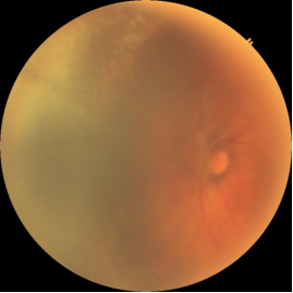
Figure 1. Case 1- Baseline.
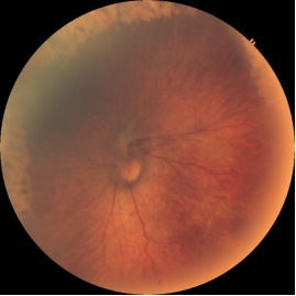
Figure 2. Case 1- At 1 month follow up.
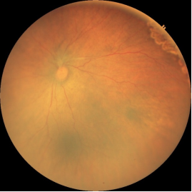
Figure 3. Case 1- At 2 months follow up.
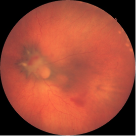
Figure 4. Case 2- Immediately following RoP Laser.
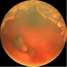
Figure 5. Case 2- One month follow-up.
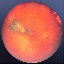
Figure 6. Case 2- Two month follow-up.
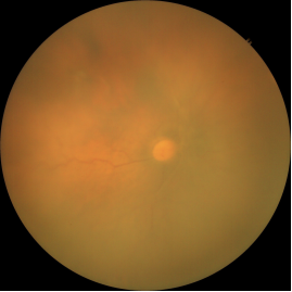
Figure 7. Case 3- Immediately following RoP Laser.
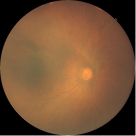
Figure 8. Case 3- At one month follow up.
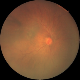
Figure 9. Case 3- At 2 month follow-up.